Visuospatial Thinking in Anatomy Education
Research Questions
Are students of an inherently high spatial ability more effective at quickly finding useful orientations of anatomical models from which to study anatomy than low spatial ability students?
If spatial ability is a factor in students ability to best use anatomical model, can we use the learning strategies of high spatial learners as a scaffold for low spatial learners in order to enhance the effectiveness of digital 3D anatomy learning tools.
Highlights
We saw significant improvement in test scores after participants were given our learning intervention
Developed a promising methodology for quantitatively understanding how students manipulate anatomical models when given a learning task.
Could not correlate the results of our chosen assessment of spatial ability with test scores.
Background
The use of computer-based 3D models in anatomy education is becoming increasingly prevalent, and yet we understand little of how students process and learn from these models. At present, no guidelines exist to maximize the educational effectiveness of these tools. With the cost of maintaining cadaver based dissections labs on the rise, it stands to reason that the use of computer based 3-D models will only increase in order to supplement anatomy curricula, thus it is ever more critical that we establish best practices for the creation of these resources . To that end it’s necessary to understand how anatomy students approach understanding spatial tasks and how to build better resources that are able to accommodate students with different visuospatial abilities while improving learning outcomes. The basis for this experiment was to see if we could improve the effectiveness of digital 3D anatomy learning tools, by using the learning strategies of high spatial learners to build a scaffold for low spatial learners, as existing digital 3D anatomy learning tools tend to leave low spatial learners at a disadvantage.

Methods
This study aimed to determine if students of an inherently high spatial ability are more effective at quickly finding useful orientations of anatomical models from which to study spatial anatomy than low spatial ability students. We did this by administering an assessment of participants spatial abilities, followed by a guided lesson in cardiac anatomy. To evaluate differences in learning performance between participants of different spatial abilities, the cardiac learning activity was preceded by a pretest and followed by a posttest. To assess participants anatomical model manipulation strategies, while completing the cardiac anatomy learning activity participants were asked to use the provided physical heart model to complete the learning task, a motion sensor embedded in the model collected orientation data relative to the models position. We hypothesized that different patterns of model manipulation between high and low spatial learners would emerge as they attempted to complete each learning task.
Participants were biology students at the University of Toronto enrolled in an introductory anatomy course. During the experiments learning intervention students were given a printed structural cardiac anatomy lesson and a physical model of the human heart, Throughout the lesson participants were asked to locate and identify specific structures on the heart model. As students worked through the lesson, we recorded how the participants were manipulating the heart model.
In order to collect this data two methods were employed:
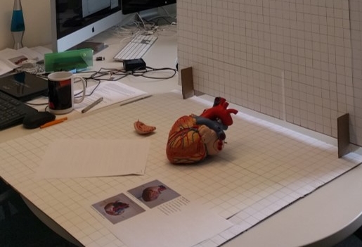
An Adafruit absolute orientation sensor (BNO 055) was embedded in the right atrium of the heart model (pictured below). This sensor wirelessly relayed x, y, and z rotation data to an Apache web server that then used a PHP script to input the data into a MySQL database. Data was relayed approximately ten times per second.
Within the test space two cameras were set up to record the participant as they worked with through the instructional package. Directly above the test area a GoPro hero camera was mounted to the ceiling, recording the participant from above. To the left of the participant a Canon T3i camera was mounted on a tripod, recording them from the left side.

Results
The basis for this experiment was to see if we could improve the effectiveness of digital 3D anatomy learning tools, by using the learning strategies of high spatial learners to build a scaffold for low spatial learners, as existing digital 3D anatomy learning tools tend to leave low spatial learners at a disadvantage. Participants showed a significant change in test score across pretest to posttest, indicating that our prepared learning package and the anatomical model were successfully able to improve the students understanding of cardiac anatomy. However, there was not a correlation between the MRT results and the pre to post test scores, rejecting our initial hypothesis.


Contributors
Dani Sayeau (Masters student)
Michael Corrin (Supervisor)
Jodie Jenkinson (Committee member)
Sanja Hinic-Frlog (Committee member)
Brian Sutherland (Arduino guru)
Andrea Gauthier (Statistics master)